We use advanced imaging and surgical technology to perform an array of surgical procedures for pets, including tracheal stenting with the aid of fluoroscopy. The trachea, or windpipe, moves air into the lungs and back out again through the nose and mouth. Rings of cartilage help the trachea stay open and maintain its shape; however, some dogs may be at risk for tracheal collapse, where the cartilage supporting the windpipe is weak or not well formed. This results in its collapse, causing the dog to cough and have difficulty breathing.
Cutting Edge Surgical can perform the necessary diagnostics to confirm tracheal collapse in dogs and perform tracheal stenting to keep the windpipe open and supported.
What is Fluoroscopy?
Fluoroscopy is an advanced imaging technique similar to X-ray, except it allows us to view areas inside the body while they are in motion. This gives us the ability to see not just the structure and appearance of an organ system, but its function as well. In cases of tracheal collapse, we can use fluoroscopy to assess the patient’s breathing ability in real time and perform a fluoroscopy-guided tracheal stenting procedure to open up the airway and mimic the function of the supporting cartilage to prevent collapse.

How Does Tracheal Stenting Work?
Tracheal stenting is a minimally invasive, non-surgical technique for treating a narrow or obstructed trachea. The tracheal stent is a self-expanding metallic device which is placed within the trachea to hold the airway open and prevent collapse. Patients must be evaluated by their veterinarian to determine whether tracheal stenting is necessary for their health and quality of life.

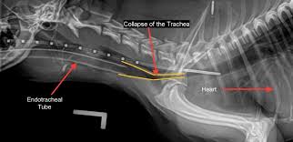
Severe tracheal collapse

Tracheal stent placement via fluoroscopy

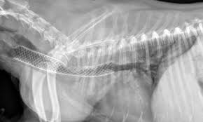
Tracheal stent in place

